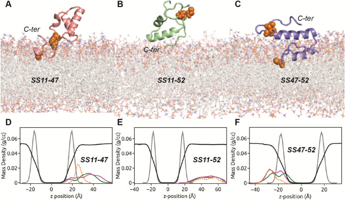
Fig. 3.
The binding modes of a SS11-47, b SS11-52 and c SS47-52 peptides with POPC bilayer are shown. The cysteine (both SS-linked and free) residues are represented as orange spheres, whilst hydrating water is not shown for clarity. The mass density profiles along the bilayer normal (z-direction) of d SS11-47, e SS11-52 and f SS47-52 peptides in POPC bilayer. The density profiles comprising hydrophobic (green) and hydrophilic (magenta), SS-bonded cysteine (orange) residues of peptide, phosphate headgroups (grey) and water (reduced by factor of 10, black) are indicated

