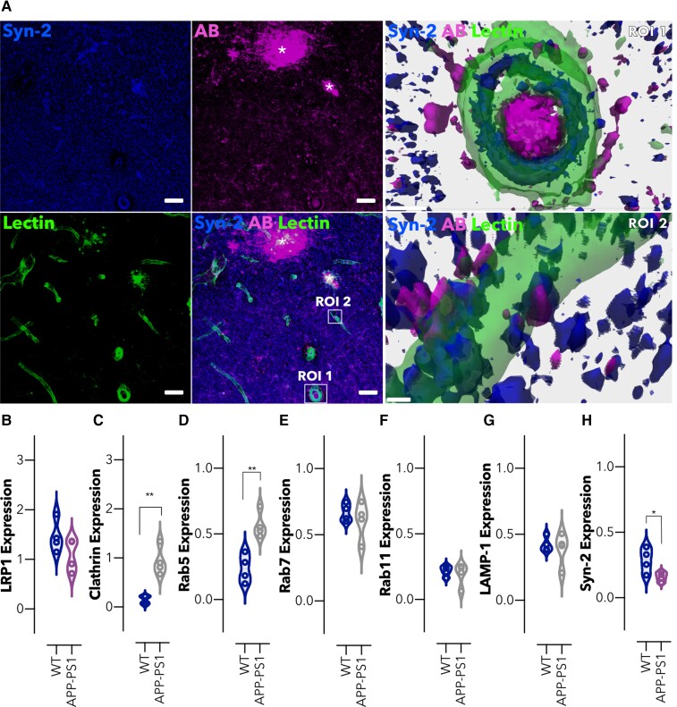
Figure 4.
Intracellular trafficking is altered in brain microvessels in an amyloidosis mouse model. (A) Representative confocal images of the hippocampal region of an Alzheimer’s disease mouse brain (APP-PS1) showing syndapin-2 (in blue), Aβ (in magenta) with lectin-labelled blood vessels (in green). Stars denote Aβ plaques colocalizing with syndapin-2. Scale bar: 20 µm. 3D reconstructions of z-stack acquired on two ROIs (ROI1 and ROI2) within the hippocampus of an APP-PS1 mouse brain exhibiting the close association of syndapin-2 and Aβ within the blood vessels. Scale bar: 5 µm. Expression levels of (B) LRP1, (C) clathrin, (D) Rab5, (E) Rab7, (F) Rab11, (G) LAMP-1 and (H) syndapin-2 in the microvessels from WT and APP-PS1 mouse brains. Data normalized to GAPDH (loading control). Mean ± SD (n = 4 animals). *P < 0.05, **P < 0.01, Student’s t-test. For full blots of the data represented in B–H, see Supplementary Fig. 9.

