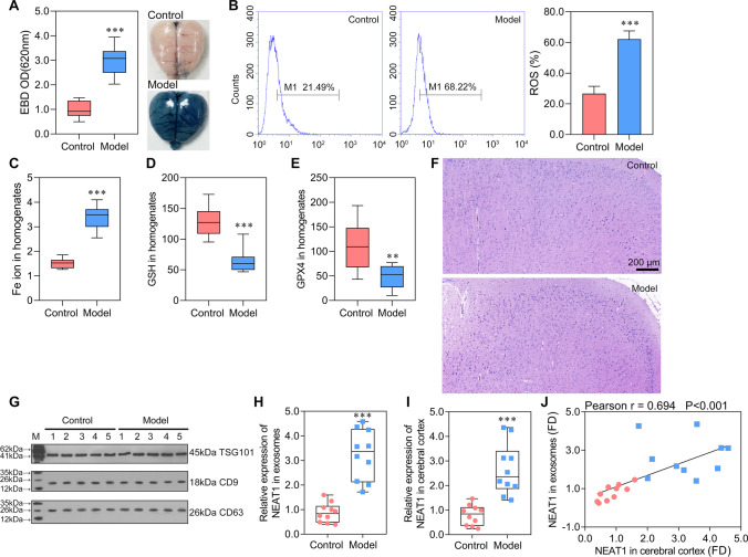
Fig. 1.
Exosome carried NEAT1 into the cerebral cortex and NEAT1 was significantly highly expressed in sepsis-induced ferroptosis A Brain vascular permeability was detected by EBD leakage in control and model rats (n = 10). Extracted dye contents in the formamide extracts were quantified at 620 nm; B ROS level in cerebral cortex homogenate was detected by flow cytometry. C–E ELISA analysis on Fe ion, GSH, and GPX4 levels. F HE staining on cerebral cortex in control and model rats. G Western blot analysis on serous exosome biomarkers of TSG01, CD9, and CD63. H, I The expression of NEAT1 was detected by qRT-PCR in the serous exosome (normalized to synthetic cel-miR-39-3p) and cerebral cortex (normalized to GAPDH), respectively. J The correlation analysis on the expression of NEAT1 in exosome and cerebral cortex. **P < 0.01 vs Control; ***P < 0.001 vs Control; EBD, Evans blue dye.

