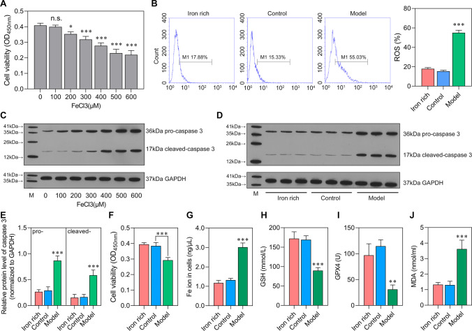
Fig. 2.
Sepsis increases stress of ferroptosis The bEnd.3 cells were stimulated by FeCl3 or serum of rats from control group and sepsis model group. A Optimal concentration of FeCl3 selection by CCK-8. n.s.: no significance; *P < 0.05, **P < 0.01, ***P < 0.001, vs 0 μM FeCl3. B ROS level in iron-rich (100 μM FeCl3), control (100 μM FeCl3 + Control serum), and model (100 μM FeCl3 + sepsis serum) group was detected by flow cytometry. C–E The protein levels of pro-caspase 3 and cleaved-caspase 3. E Cell viability was detected using CCK-8. F–I The levels of Fe ion (Fe2+ and Fe3+), GSH, GPX4, and MDA. *P < 0.05, **P < 0.01, ***P < 0.001 vs iron-rich and control.

