Fig. 3.
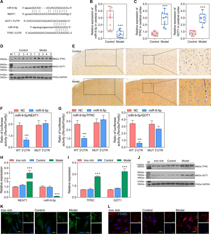
miR-9-5p, sponged by NEAT1, overexpressed in vitro and in vivo and might promote cell apoptosis in sepsis-induced ferroptosis by targeting TFRC and GOT1 A The binding regions between miR-9-5p with sequences from NEAT1, TFRC, and GOT1. B, C The expression level of miR-9-5p, TFRC, and GOT1 in model and control was tested by qRT-PCR in vivo (normalized to U6 or GAPDH). ***P < 0.001 vs control. D The protein levels of TFRC and GOT1 in cerebral cortex were assessed using Western blotting. E The protein level of TFRC in cerebral cortex was showed by immunohistochemistry. F, G The dual-luciferase reporter gene assay analyzes the binding relationship between miR-9-5p with sequences from NEAT1, TFRC, and GOT1. ***P < 0.001 vs NC. H, I The expression level of miR-9-5p, TFRC, and GOT1 in iron-rich (100 μM FeCl3), control (100 μM FeCl3 + control serum), and model (100 μM FeCl3 + sepsis serum) group was tested by qRT-PCR (normalized to U6 or GAPDH). *P < 0.05, **P < 0.01, ***P < 0.001 vs iron-rich and control. J The protein levels of TFRC and GOT1 assessed using western blotting in vitro. K, L The immunofluorescence for detecting TFRC and GOT1 (blue, DAPI; green, TFRC; red, GOT1).

