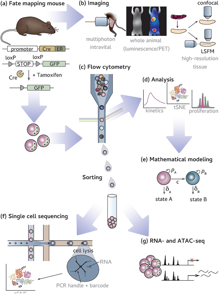
Figure 3. Examples of experimental techniques that are compatible with fate-mapping mice.
(a) Fate-mapping begins with the temporal expression of a reporter allows for fate-mapping. Following fate-mapping, researchers can use many tools to understand how cellular origin can dictate phenotype and function. (b) Imaging technologies are a powerful collection of tools to assess in situ localization and interactions with other cell types. (left) Multiphoton imaging is used to perform intravital imaging deep into animal tissues of interest [121] (an example is live imaging of cellular interactions and dynamics in lymph nodes); (middle) whole animal imaging, either by luminescence or positron emission tomography (PET) modalities, allows for non-invasive longitudinal study; (right) advances in tissue clearance (e.g. 3DISCO) [122] improve high resolution microscopy of tissues with techniques such as confocal and light sheet fluorescent microscopy (LSFM). (c) Flow cytometry uses laser excitation to measure fluorescence of labeled markers on cells. (d) Traditional and high parameter analyses can be performed via flow cytometry data. (e) Mathematical modeling uses accumulated data to model cellular behavior. Flow cytometry can also sort cells, based on specific marker expression, for a variety of downstream technologies such as (f) single cell sequencing analysis in which an individual cell’s transcripts are associated with a unique barcode and (e) bulk RNA and ATAC sequencing in which cellular populations are sequences for transcriptional (RNA) or chromatin accessibility (ATAC) information [94,129].

