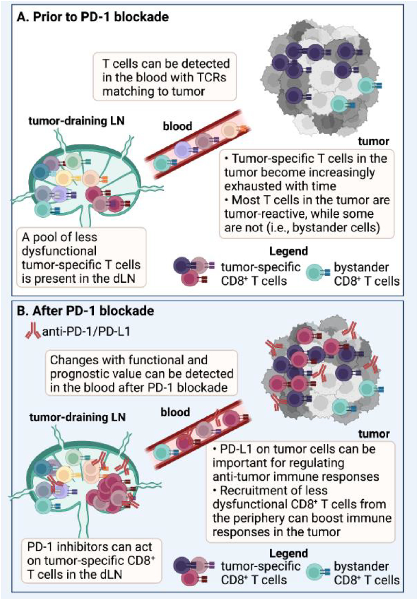
Figure 2: Use of T cell receptor (TCR) sequencing to inform mechanisms of PD-1-based immunotherapy.

(A) The TCR sequence has been used as a molecular barcode to track T cells with shared TCRs between the tumor-draining lymph node (LN), blood, and tumors in mice [27, 63]. Similarly, the TCR has been used as a barcode to track T cells with matching TCRs between blood and tumor in humans [27–29, 35]. The tumor-draining LN contains a reservoir of less dysfunctional T cells, while the T cells found in the tumor often display a more exhausted or dysfunctional phenotype [31, 56, 63]. In addition to tumor-specific T cell clones, bystander T cells can also be present in the tumor that are not specific for canonical tumor antigens [107], but may still contribute functionally to the anti-tumor response [108, 109]. (B) Work has shown that Programmed Death 1 (PD-1) pathway inhibitors can act on T cells in the tumor-draining LN in mice [61, 62], and immunological changes that have functional and prognostic significance can be detected in the peripheral blood of humans [40, 67, 131–133]. An important mechanism of protection following PD-1 pathway blockade is recruitment of new, potentially less dysfunctional T cell clones from the periphery into the tumor [30, 36, 38–40, 67]. However, studies in mice and humans suggest that PD-1 inhibitors also likely act on T cells in the tumor as well [56–59]. Abbreviations in Figure: dLN = draining lymph node; TCR = T cell receptor, PD-1 = Programmed Death 1. Created with BioRender.com.
