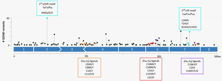
Figure 2.
Diagram of the coding SDHB variants along the amino acid sequence L(I)YR motifs are shown in blue. The cysteine residues ligating the [Fe2-S2], the [Fe4-S4] and the [Fe3-S4] are shown in orange, red and purple, respectively. Diagram is displayed as lollipop symbols designed using the Mutation Mapper tool of the cBioPortal website. The Y axis represents the number of occurrences of variant in one residue.

