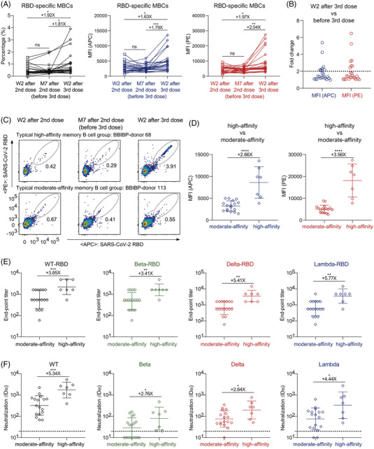
FIGURE 3.

High‐affinity RBD‐specific memory B cells elicited by a third dose of inactivated vaccine. (A) The percentage (left), mean fluorescence intensity (MFI) of APC (middle), and MFI of PE (right) of RBD‐specific MBCs (CD19+CD3−CD8−CD14−CD27+IgG+SARS‐CoV‐2‐RBD+ cells) of randomly selected 24 donors with three follow‐up visits. (B) The fold change of MFI in both APC and PE of RBD‐specific MBCs between week 2 after third vaccination and before third vaccination. A cut‐off of twofold is indicated by the horizontal dashed line. High‐affinity group: fold change >2, moderate‐affinity group: fold change <2. (C) The typical display of high‐affinity and moderate‐affinity RBD‐specific MBCs of two donors with three follow‐up visits (high: BBIBP‐donor 68, moderate: BBIBP‐donor 113). (D) Comparison of MFI in both APC (left) and PE (right) of RBD‐specific MBCs at week 2 after third vaccination between the high‐affinity group (n = 7) and the moderate‐affinity group (n = 17). (E) The end‐point titres of binding IgG to SARS‐CoV‐2 WT, beta, delta, and lambda RBD proteins at week 2 after third vaccination in the high‐affinity and moderate‐affinity groups. (F) The geometric mean titres of nAbs against SARS‐CoV‐2 WT, beta, delta, and lambda pseudoviruses at week 2 after third vaccination in the high‐affinity and moderate‐affinity groups. A cut‐off of 1:20 dilution is indicated by a horizontal dashed line. ID50, 50% inhibitory dilution. ‘+’ indicates an increase. ‘X’ indicates fold change. *p < .05; **p < .01; ***p < .001; ****p < .0001; ns, not significant
