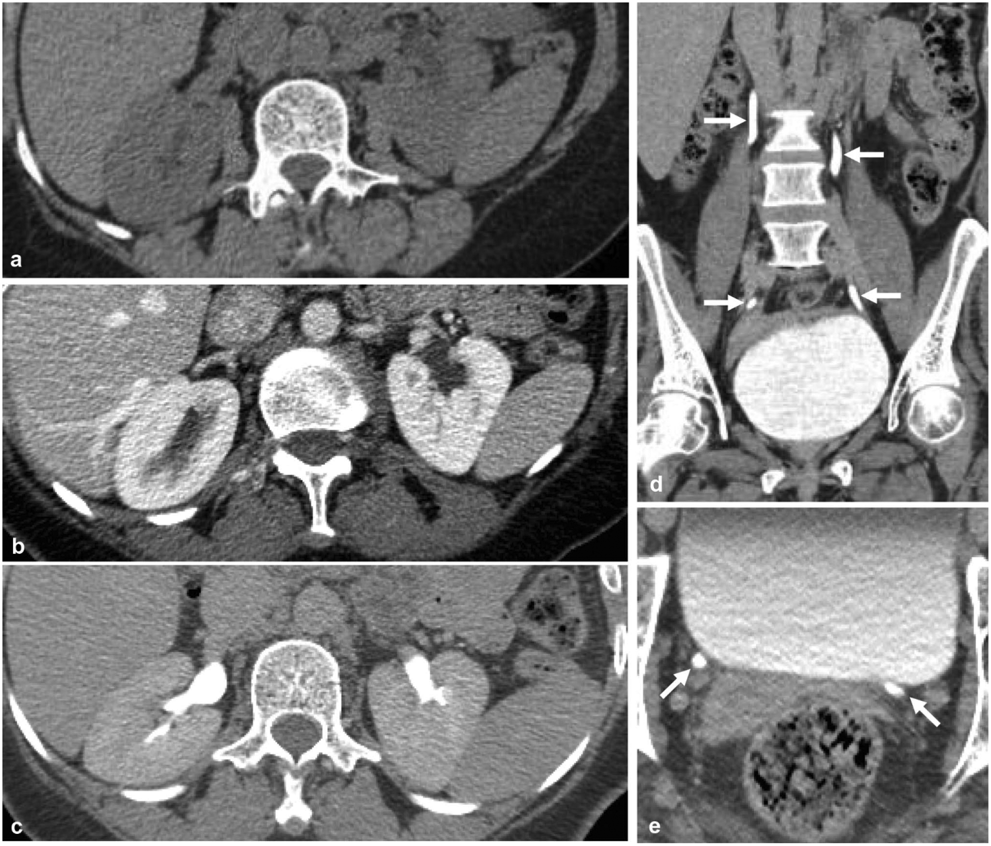
Fig. 1.

Normal appearance of the three phases of CT urography is provided for reference. a Non-contrast axial image through the kidneys shows no renal calcifications or high attenuation masses. b Nephrographic phase axial image through the kidneys show symmetric parenchymal enhancement without parenchymal mass. c Excretory phase axial image through the kidneys shows symmetric excreted contrast in bilateral renal pelvises. Symmetric contrast opacification of the ureters (arrows) to the level of the bladder, as shown on d the coronal reformatted excretory phase image and e the excretory phase axial image at the level of the ureteral insertion to the bladder
