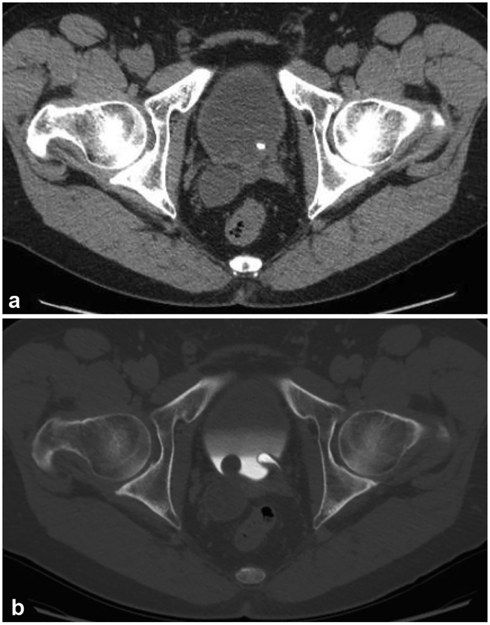
Fig. 11.

A 47-year-old male with incidental finding of right-sided hydroureter on imaging for work-up of an unrelated issue. a Non-contrast axial image of the pelvis demonstrates right distal hydroureter. There is also a high density in the bladder in the region of the left ureterovesicular junction (UVJ). b Excretory phase axial image on bone windows demonstrates bilateral ureteroceles, non-opacified on the right due to impaired excretion by the right kidney. The classic “spring onion” sign is best demonstrated on the left where the ureterocele is opacified with contrast. The left UVJ calculus seen on the non-contrast image is shown to be within the left ureterocele. The ureterocele on the right is likely the cause of this patient’s right-sided hydroureter
