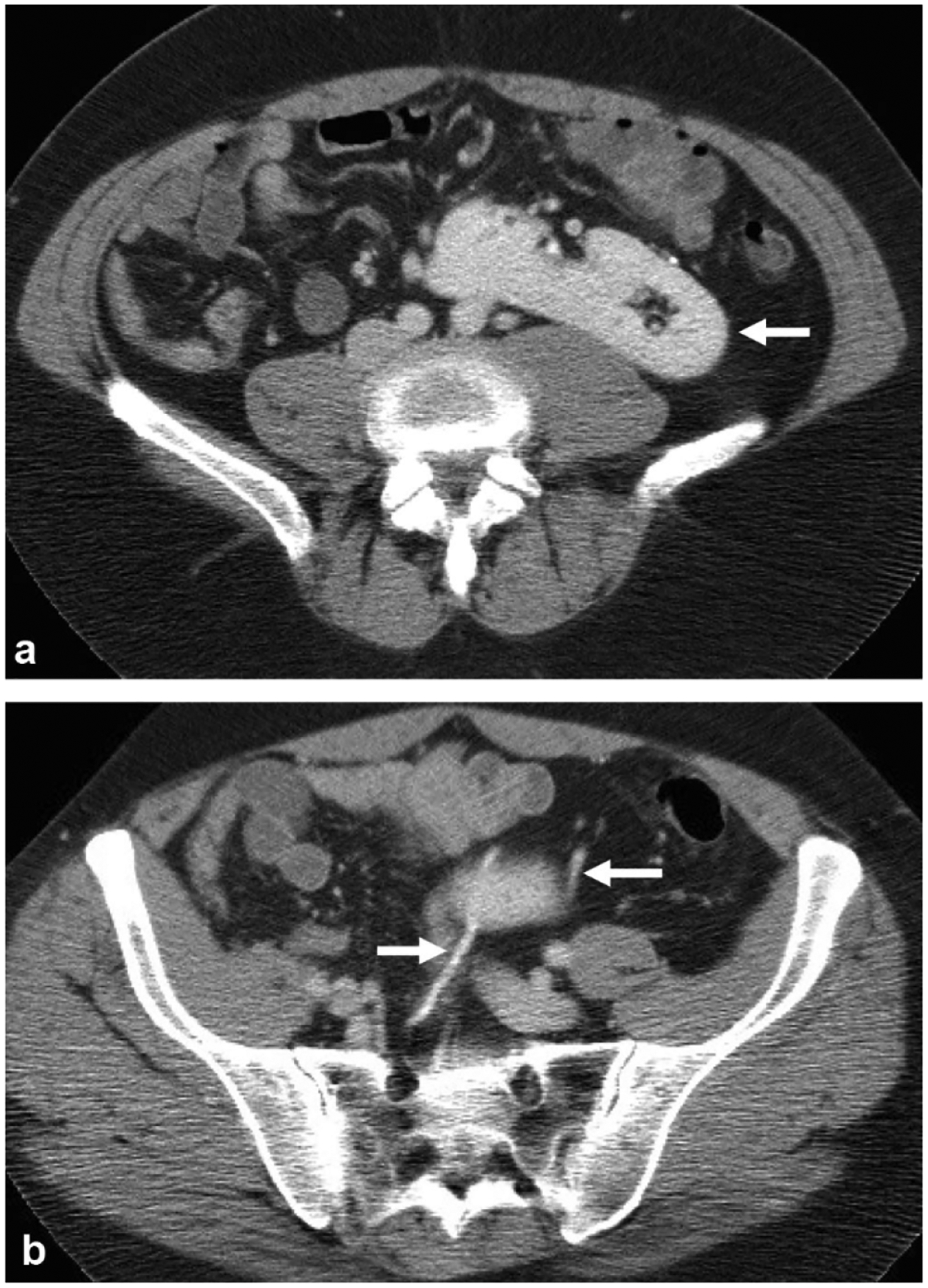
Fig. 14.

A 46-year-old female presents with abdominal pain, incidentally found to have crossed fused renal ectopia. a Nephrographic phase axial image at the level of the kidneys shows an ectopic right kidney fused to the left kidney (arrow) in the left upper quadrant. b Excretory phase axial image shows two ureters (arrow), one of which crosses midline to the right hemiabdomen before inserting on the bladder at the right UVJ
