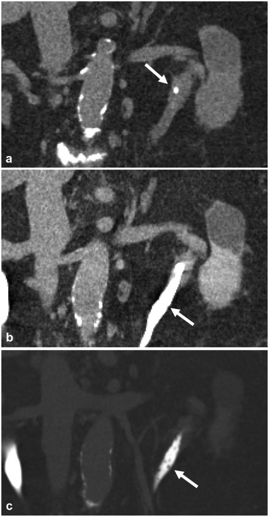
Fig. 15.

A 72-year-old female with chronic UTI. a Non-contrast coronal reformatted image demonstrates a ureteral stone (arrow) in the left proximal ureter. b Excretory phase coronal reformatted image shows very subtle indentations (arrow) in the proximal and mid ureter. c The same coronal reformatted image in bone windows reveals multiple uniform tiny filling defects (arrow) in the proximal and mid ureter, which represent multiple subepithelial cysts in the wall of the ureter from ureteritis cystica
