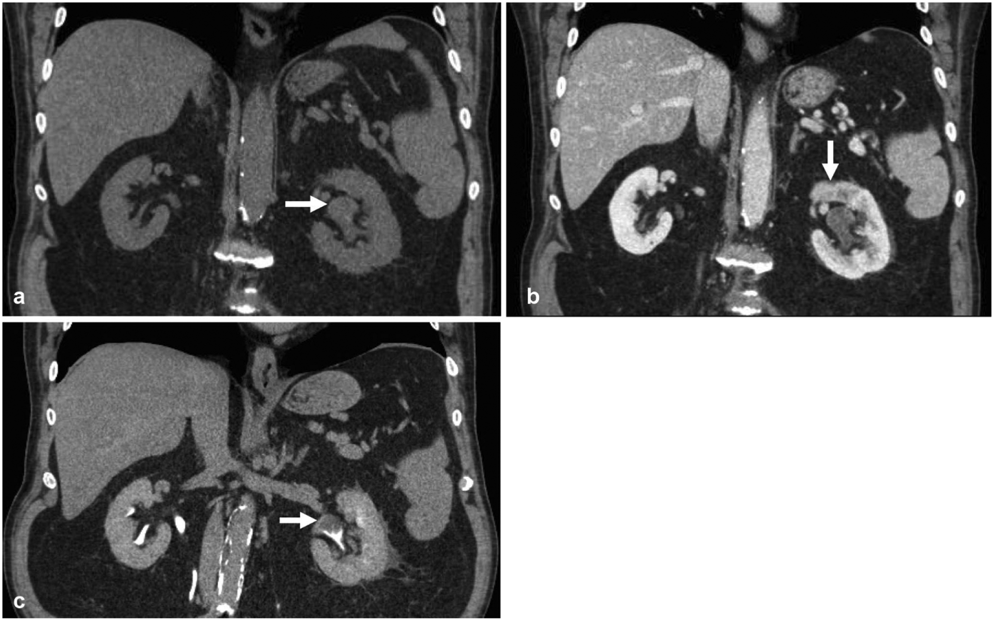
Fig. 2.

CT urogram was obtained in this 63-year-old male for evaluation of gross hematuria. a Non-contrast coronal reformatted image shows mild left pelviectasis (arrow). b Nephrographic phase coronal reformatted image shows subtle heterogeneous enhancement in the left renal pelvis (arrow) and delayed left nephrogram relative to the right. c Excretory phase coronal reformatted image shows a clear filling defect within the renal pelvis (arrow), which was later confirmed as urothelial carcinoma
