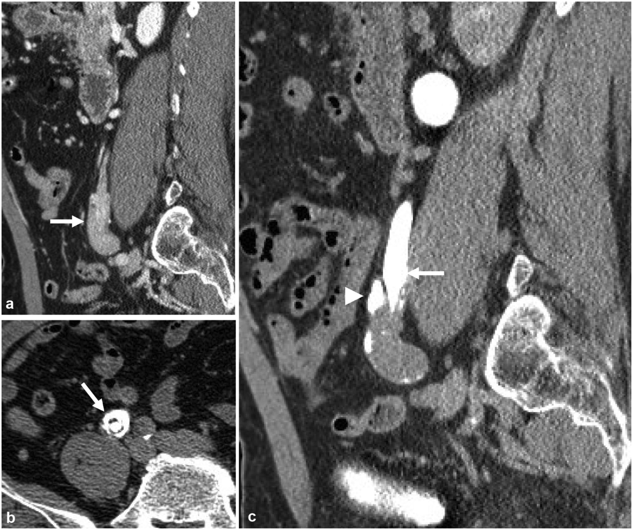
Fig. 5.

A 65-year-old male with microhematuria and flank pain. a Nephrographic phase sagittal reformatted image demonstrates bulbous appearing mass at the distal right ureter (arrow). b Excretory phase axial image better demonstrates contrast circumferentially in a “target sign” appearance suggestive of ureteroureteral intussusception (arrow), which was due to a urothelial carcinoma as a lead point. c Excretory phase sagittal reformatted image demonstrates tapering of the distal ureter, or the intussusceptum (arrow), with contrast seen peripherally, also called the intussuscipiens (arrowhead)
