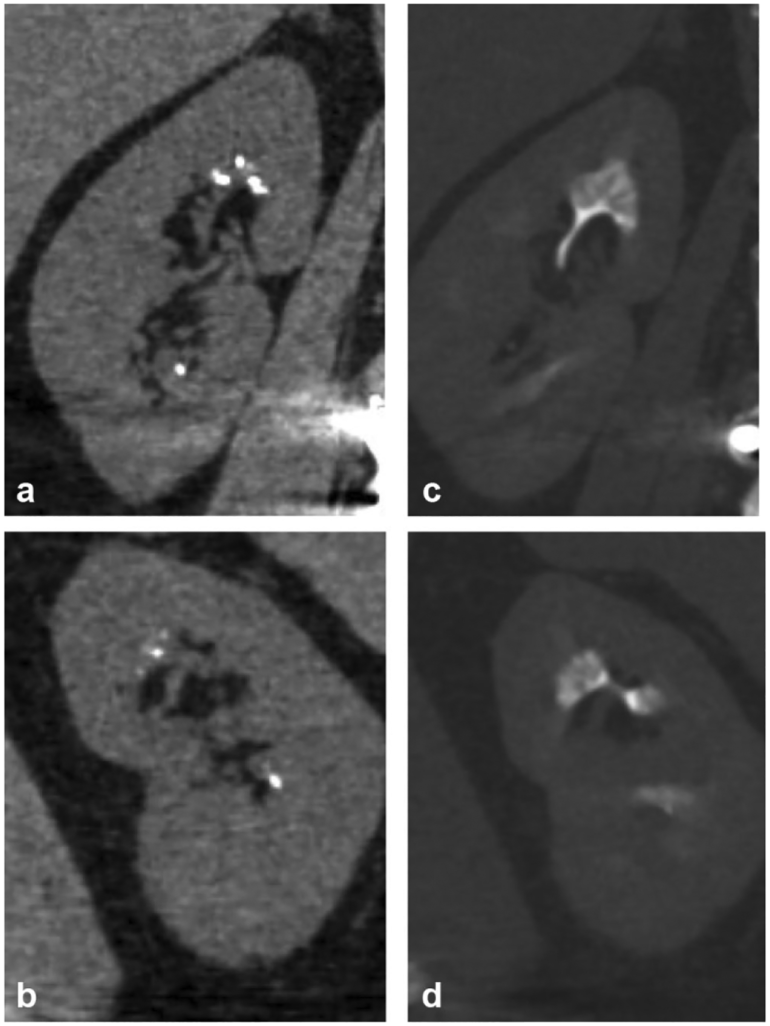
Fig. 6.

A 63-year-old male undergoing work-up for renal insufficiency. Non-contrast coronal reformatted images of the left (a) and right (b) kidneys demonstrate medullary calcifications in bilateral kidneys that may easily be mistaken for small renal calculi. Excretory phase coronal reformatted images in bone windows of the left (c) and right (d) kidneys demonstrate the characteristic “paintbrush appearance” of medullary sponge kidney, presumably from contrast filling-dilated medullary collecting ducts
