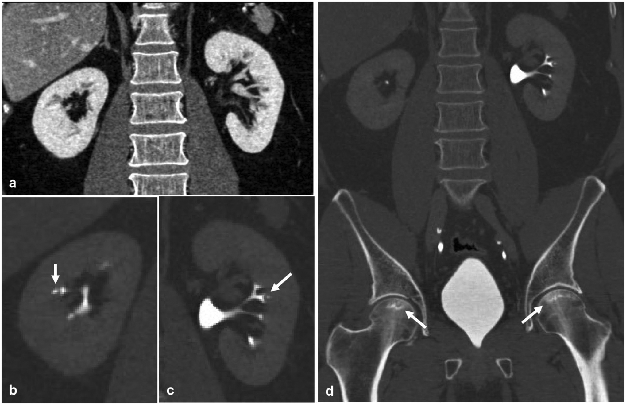
Fig. 7.

A 29-year-old male with sickle cell disease presents with bilateral flank pain. a Nephrographic phase coronal reformatted image of the kidney demonstrates an otherwise normal appearing symmetric nephrogram. Excretory phase coronal reformatted images in bone windows of the b right and c left kidneys demonstrate the classic “golf ball on tee” sign (arrow) of renal papillary necrosis. d Bilateral femoral head osteonecrosis (arrows) is also seen, which in the absence of the given history would help solidify sickle cell disease as the etiology behind this patient’s renal papillary necrosis
