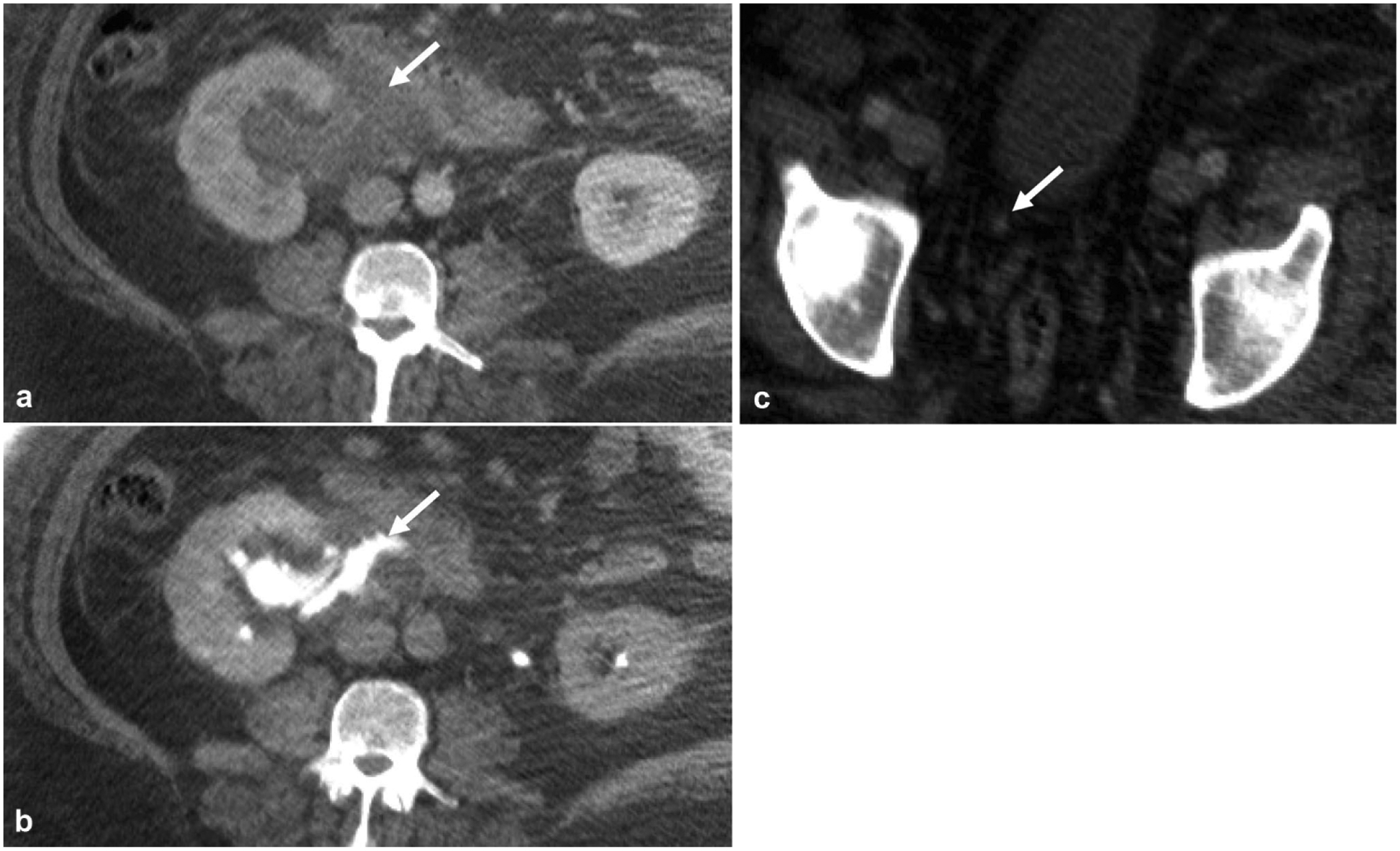
Fig. 9.

A 50-year-old male with right flank pain that acutely improved before CT exam. a Nephrographic phase axial image shows a right peripelvic fluid collection. b Excretory phase axial image shows contrast extending into the fluid collection from the renal pelvis compatible with a urinoma from forniceal rupture, which in this case was confirmed to be secondary to a c 2-mm obstructing distal ureteral stone (arrow)
