Figure 6.
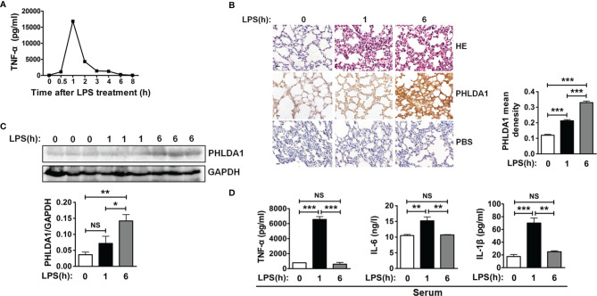
PHLDA1 alleviates LPS-initiated proinflammatory cytokine production in a mouse model of endotoxemia. (A) ELISA was performed to measure TNF-α level in serum from mice treated with LPS (8 mg/kg) for 0, 0.5, 1, 2, 3, 4, 6, and 8 h, respectively. (B) H&E and PHLDA1 staining (scale bar, 20 μm) of lung tissues from three groups of mice, including group 1 (control group), group 2 (treatment with LPS for 1 h), and group 3 (treatment with LPS for 6 h). PHLDA1 mean density was analyzed and shown in the right panel. (C) Western bolt analysis of PHLDA1 in the lung tissues from the above different groups of mice. The quantified result of PHLDA1 expression is shown in the lower panel. (D) ELISA analysis of proinflammatory cytokines (TNF-α, IL-6, and IL-1β) in serum from the above different groups of mice. The data, including quantitative analysis of PHLDA1 expression, PHLDA1 mean density, and ELISA, are expressed as mean ± SD from three independent experiments. (NS means no significance, *P < 0.05; **P < 0.01; ***P < 0.001).

