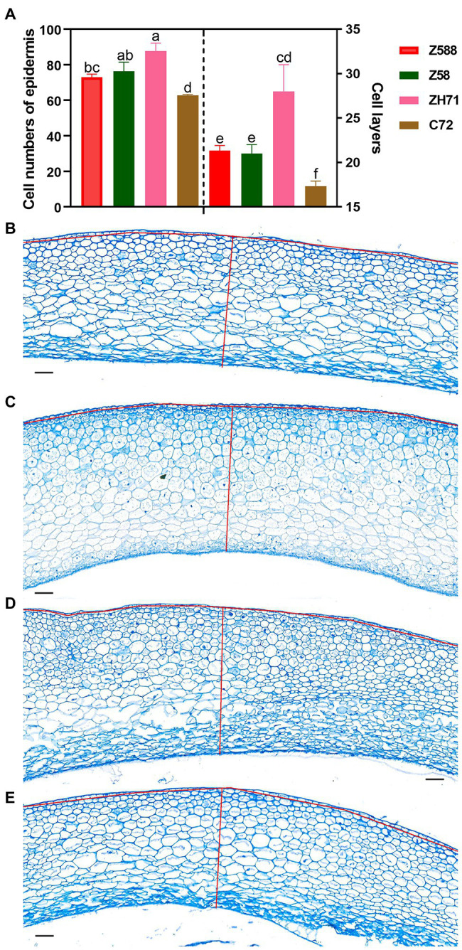
Figure 6.

Cell division of the kernel pellicle of the four inbred lines at early seed developmental stage. (A) To determine the cell number and number of cell layers in the kernel pellicle, kernels were collected from plants that grown in three different blocks with three replicates per genotype (Z588, Zheng588; ZH71, ZhengH71; Z58, Zheng58; and C72, Chang7-2) per block at 10 d after pollination. Each value is shown as the average of six independent experiments ± SD. Different letters indicate significant difference (p < 0.05) as determined by Tukey–Kramer test. Micrographs of the kernel pellicle from Z588 (B), Z58 (C), ZH71 (D), and C72 (E). For estimation of the numbers of cells and layers, the number of cells along the red line were scored. The scale bar represents 50 μm.
