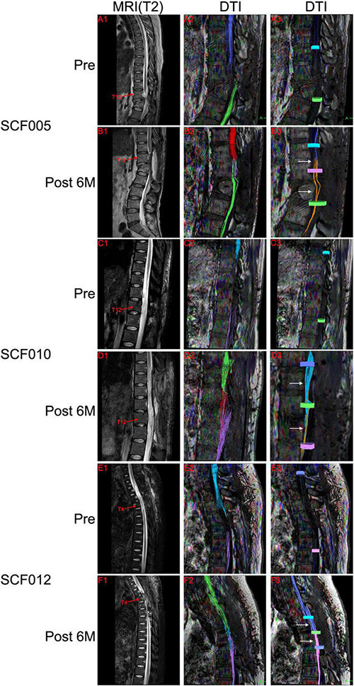
FIGURE 8.

Representative neuroimages. Preoperative T2-weighted MRI scans showed spinal cord injury (A1,C1,E1), and DTI showed complete disruption of the spinal cord fibers (A2,A3,C2,C3,E2,E3). Postoperative T2-weighted MRI scans and DTI showed the autotransplanted sural nerve in the original spinal cord injury area (B1,B2,D1,D2,F1,F2). DTI showed restoration of neural connection at the two sites of transection (white arrows in B3,D3,F3). The colors of the fibers were automatically generated by the DTI system of MRI, with no practical significance. Pre, Preoperatively; Post 6M, 6 months postoperatively.
