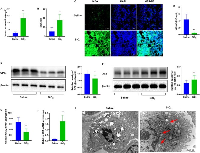
Figure 2.

SiO2 induces ferroptosis in mice. Mice were intratracheally injected with SiO2 (100 mg/ml) for 56 days. n = 6 per group. (A) Iron concentration in the lung tissue sections from the indicated groups. (B) Malondialdehyde (MDA) levels for peroxide formation and (C) MDA staining. Scale bars, 50 μm. (D) The glutathione/glutathione oxidized (GSH/GSSG) assay. Western blotting and quantification of the (E) GPX4 and (F) XCT proteins in lung tissues from saline and SiO2 groups. mRNA expression of (G) GPX4 and (H) XCT in lung tissues from mice exposed to saline or SiO2. (I) Mitochondrial morphological changes were detected via transmission electron microscopy (TEM). Typical mitochondrial morphology in ferroptosis is indicated with red arrows. Scale bars, 1000 nm. *P < 0.05 vs. saline group; **P < 0.01 vs. saline group.
