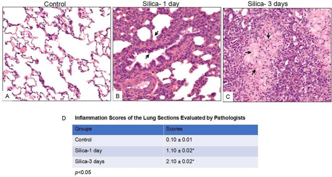
Figure 1.
Histological analysis of the lung tissues from silica-exposed rats compared with controls following intratracheal instillation (IT) for 1 day and 3 days. Rats were received IT exposure to silica (50mg/mL, 1mL) or sterile saline. One day and 3 days following exposure, the animals were sacrificed and lung tissues were processed for histochemical analysis. The control animals were sacrificed one day after saline IT exposure. A comparison of the gross architecture of the lung sections from saline-exposed rats revealed normal lung structure (A). In contrast, silica-exposed rats developed features of pulmonary inflammation and edema, characteristic of rat silicosis (B & C). (D) Inflammation score of rat lung tissue was evaluated by two pathologists in a double-blind fashion, taking no inflammation as 0 score, mild inflammation as 1, overt leukocyte infiltration as 2, and massive infiltration as 3. The images were reprehensive pictures from 5 animals of each group. 200⨯ original magnification. *p<0.05.

