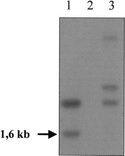
Figure 3.
Southern-blot analysis. The genomic DNAs from reference strains were digested by PstI and separated by electrophoresis. The hybridization patterns were obtained with a 323-bp probe corresponding to the bacterial part of the pARG7 plasmid that was used for mutagenesis. The original sta8-1::ARG7 mutant strain (BafV13) profile is displayed in lane 1. The wild-type strain profile 330 used for the insertional mutagenesis (lane 2) shows no signal. Strain BafO6 (sta8-2::ARG7) profile is displayed in lane 3. The arrow corresponds to the 1.6-kb signal cosegregating with the sta8-1::ARG7 mutation.

