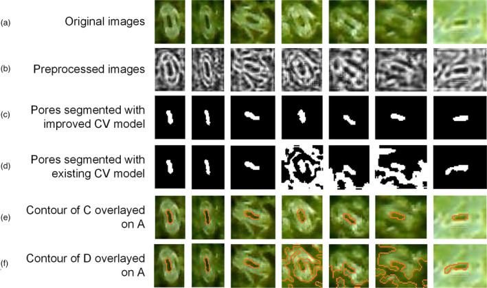
Figure 8.

Pore segmentation of a single stoma image of the improved CV model vs Li’s CV model. (a) Original stoma images. (b) Preprocessed grey‐level image obtained with the Lucy–Richardson algorithm and CLAHE. (c) The pores segmented with the improved CV model. (d) The pores segmented with existing CV model. (e) The contour of C overlayed on the original images. (f) The contour of D overlayed on the original images.
