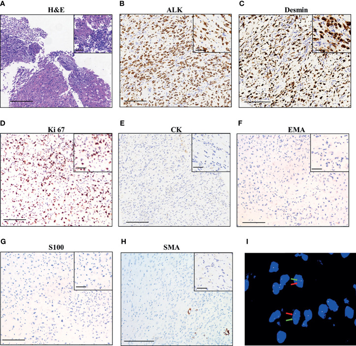
Figure 1.
Pathological findings of the patient. (A) Hematoxylin–eosin staining of surgical tumor sample. IHC staining of ALK (B), desmin (C), Ki 67 (D), CK (Cytokeratin, (E), EMA (F), S100 (G), and SMA (H). The magnification in (A–H) is 100×, scale bar is 200 μm; the magnification in the inset images is 400×, scale bar is 50 μm. (I) Fluorescence in situ hybridization (FISH) using a break-apart ALK locus probe. ALK gene rearrangement is indicated by the split signals (indicated by red and green arrows, magnification 1000×).

