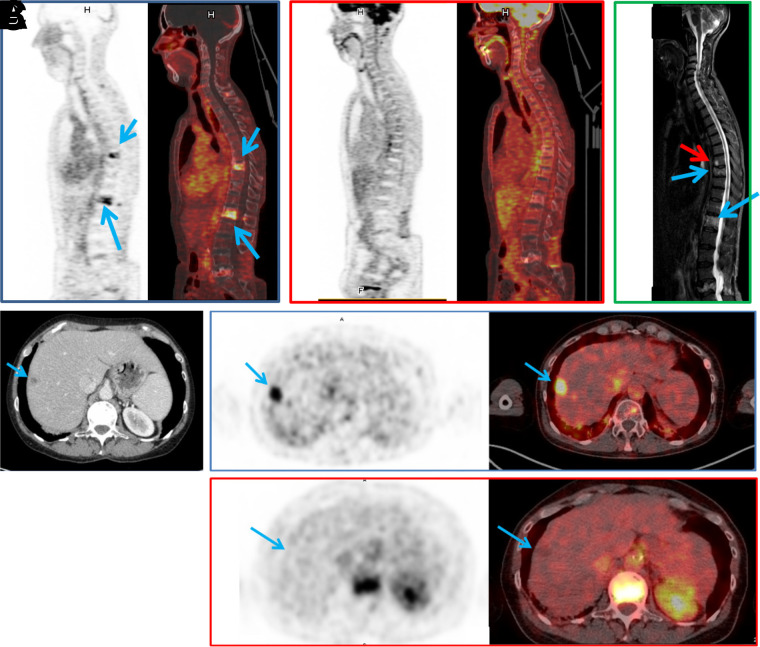
FIGURE 3.
(A–C) In patient 1, pretargeted immuno-PET with TF2 and 68Ga-IMP288 peptide images show 2 vertebral metastases (L1 and T9, arrows) (A), 18F-FDG PET discloses no vertebral abnormalities (B), and vertebral MRI confirms both lesions (blue arrows) and discloses another lesion (red arrow) at T8 (C). (D–F) In patient 2, CT shows suspected liver lesion (D), and pretargeted immuno-PET with TF2 and 68Ga-IMP288 peptide reveals high uptake by liver lesion (arrow) (E), which was not seen by 18F-FDG PET (F). (Reprinted from (21).)

