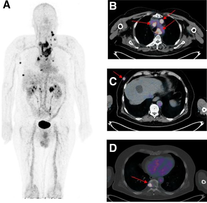
FIGURE 1.

Immuno-PET/CT with anti-CEA BsmAb and 68Ga-IMP288 peptide showing pathological lesions with heterogeneous SUVmax ranging from 3.0 to 20.1. Maximum-intensity-projection (MIP) image (A) showed several pathological lesions. On the fusion axial images, arrows located mediastinal nodes (B), subcutaneous lesions (C), and bone metastasis (D).
