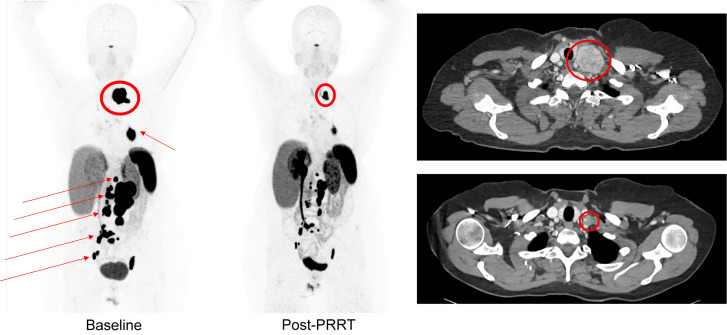
FIGURE 3.
A 53-y-old woman with metastatic hormone-secreting SDHB-associated pheochromocytoma. Maximal-intensity-projection images of 68Ga-DOTATATE at baseline (left) vs. 12 mo after PRRT with 7.4 GBq of 177Lu-DOTATATE 4 times (middle) demonstrate significant decrease in tracer uptake in tumors in neck (encircled), left hilum, retroperitoneum, and pelvis (arrows). Baseline axial CT image (top right) through neck demonstrates large left supraclavicular mass at baseline (encircled), and post-PRRT axial CT image (bottom right) shows significant decrease in tumor size (encircled).

