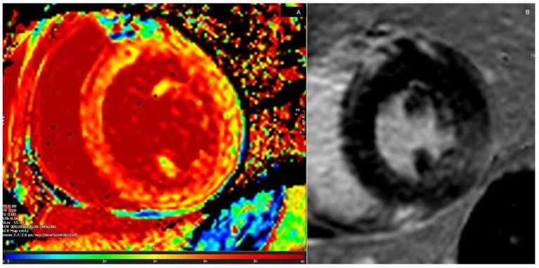
Figure 2.
Global extracellular volume by cardiovascular magnetic resonance imaging of Case 1. Pathological values were obtained from the entire left ventricular circumference (A). Focal conspicuities were shown in late gadolinium enhancement (LGE) sequences. This showed inferolateral LGE consistent with regional wall motion abnormalities on echocardiography (B).

