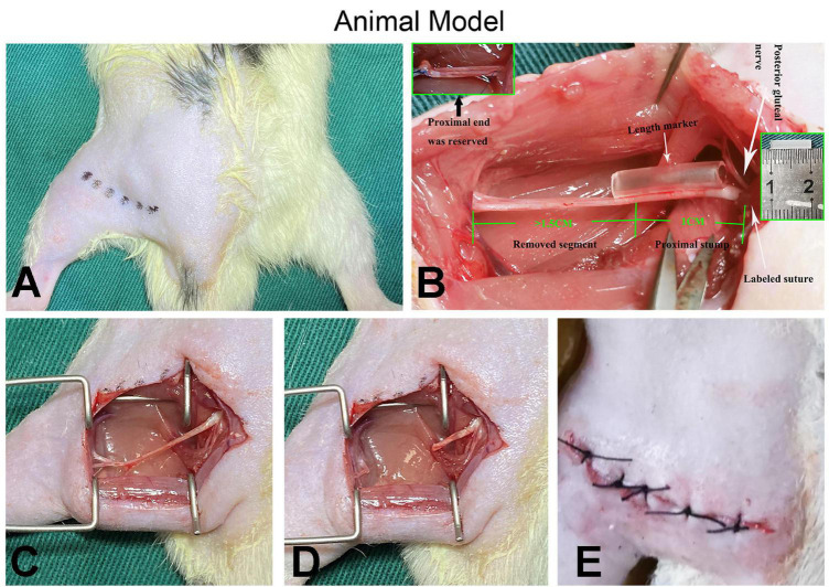
FIGURE 2.
Demonstration of surgical procedures. (A) After the animal is shaved, the surgical entry position (the muscle gap between the biceps femoris and the gluteal muscles) is marked, the surgical area is disinfected, and draped. (B) The white arrow on the right indicates the origin of the posterior gluteal nerve (top) and the location where the marked sutures are placed for quantitative analysis (bottom); the white arrow in the middle indicates the length marker (silicone catheter in the middle) for precise nerve cutting. The green numbers 1 and 1.5 and the illustrations (top left and middle right) show the details of the preparation of the proximal nerve stump. (C–E) The muscle wound and the skin incision are sutured with 4-0 sutures after the preparation process and completion of the PNS (proximal nerve stump).

