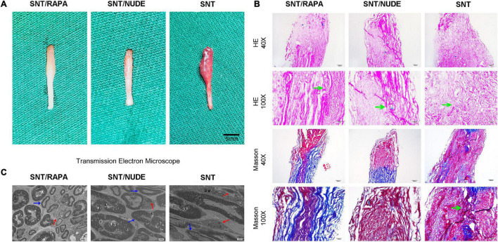
FIGURE 4.
The results of gross observation and histological analysis of the PNS. (A) Images of neuromas in all groups. Typical globular neuromas were observed in both the SNT group and the SNT/RAPA group 30 days after the neurectomy, but no such findings were observed in the SNT/NUDE group. (B) H&E and Masson’s staining of neuroma 30 days after surgery. H&E staining of traumatic neuroma in the SNT/NUDE group under the light microscope, showing that the number of fibroblasts was extremely small, while the SNT/RAPA group and SNT group had a large number of fibroblasts (green arrows). Masson’s staining shows that in the SNT/NUDE group the collagen is slightly blue, and the nerve fibers are arranged relatively orderly. In the SNT group, the dense blue-stained collagen has randomly arranged nerve bundles, a large number of axons in neuromas, and more spindle fibroblasts and collagen fibers (as shown by the green arrow). Compared to the SNT group, there are fewer spindle fibroblasts and collagen fibers in the SNT/RAPA group, showing dark blue stained collagen, with regularly arranged nerve bundles. (40×, scale bar: 200um; 100×, scale bar 100 um) (C) Transmission electron microscopy was generally observed 30 days post-surgery. SNT/NUDE group: There are a large number of thick myelinated fibers with few fibroblasts; the red arrows indicate scattered unmyelinated fibers, and the blue arrows indicate myelinated fibers; only a few collagen fibers can be seen at random. SNT group: There are a large number of unmyelinated fibers and abundant fibroblasts; abundant horizontal and diagonal collagen fibers are randomly distributed; the marks show dense horizontal collagen fibers (red arrow) and myelin sheath (blue arrow). SNT/RAPA group: collagen fibers are distributed at the bottom; blue arrows indicate Schwann cells of internal mitochondria and endoplasmic reticulum, and red arrows indicate unmyelinated nerve fibers. (Magnification 3000×).

