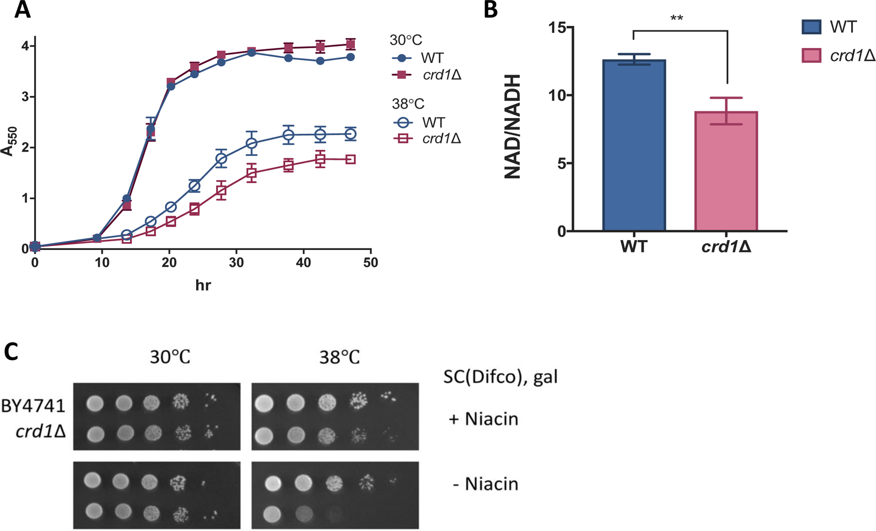
Figure 1. The NAD(H) redox balance is disrupted in crd1Δ.

(A) Precultures in the stationary phase were inoculated to a concentration of A550=0.05 in fresh SC-galactose medium and incubated with aeration. A550 was measured at indicated times. Data shown are mean ± S.D. (n=3). (B) The NAD+/NADH ratio was measured in cells grown in galactose medium to the mid-logarithmic phase as described in Materials and Methods. Data shown are mean ± S.D. (n=3). Unpaired t test, p=0.0033 (C) 10-fold serial dilutions of WT and crd1Δ cells spotted on SC-galactose medium with or without niacin. Plates were incubated at 38.5 ± 0.5 °C for 4 days.
