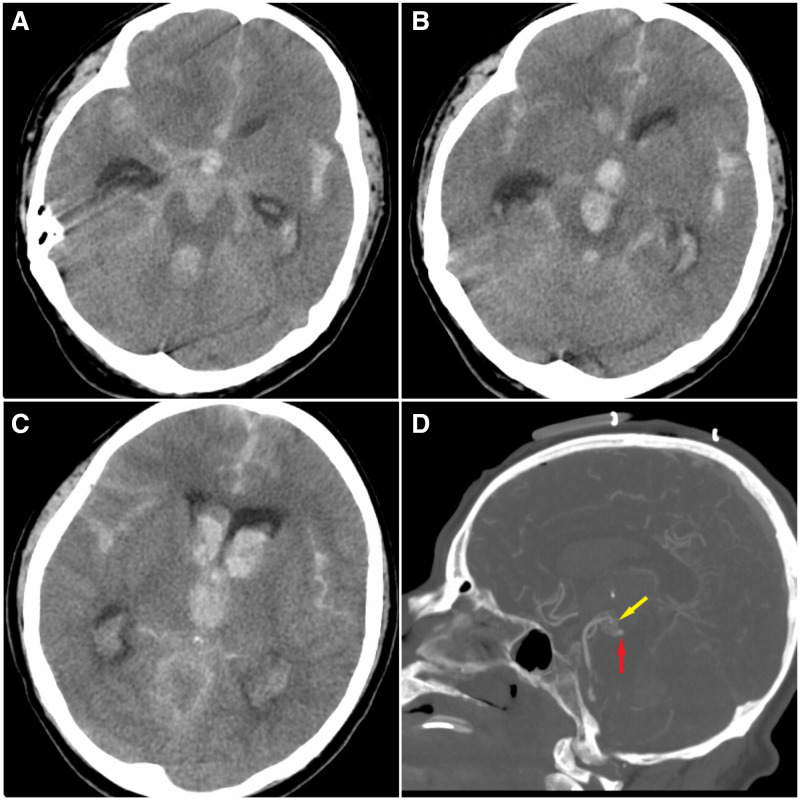
Figure 1.
(A) Non-enhanced CT scan revealing severe acute cisternal subarachnoid haemorrhage, especially in the interhemispheric and sylvian compartments, as well as acute brainstem intraparenchymal haemorrhage. (B, C) More rostral slices of the same CT scan demonstrating intraventricular haemorrhage in the third ventricle and frontal horn of the lateral ventricles. (D) CT angiography demonstrating an abnormal vascular network at the tip of the basilar artery and possibly a small aneurysm.

