
Fig. 7.
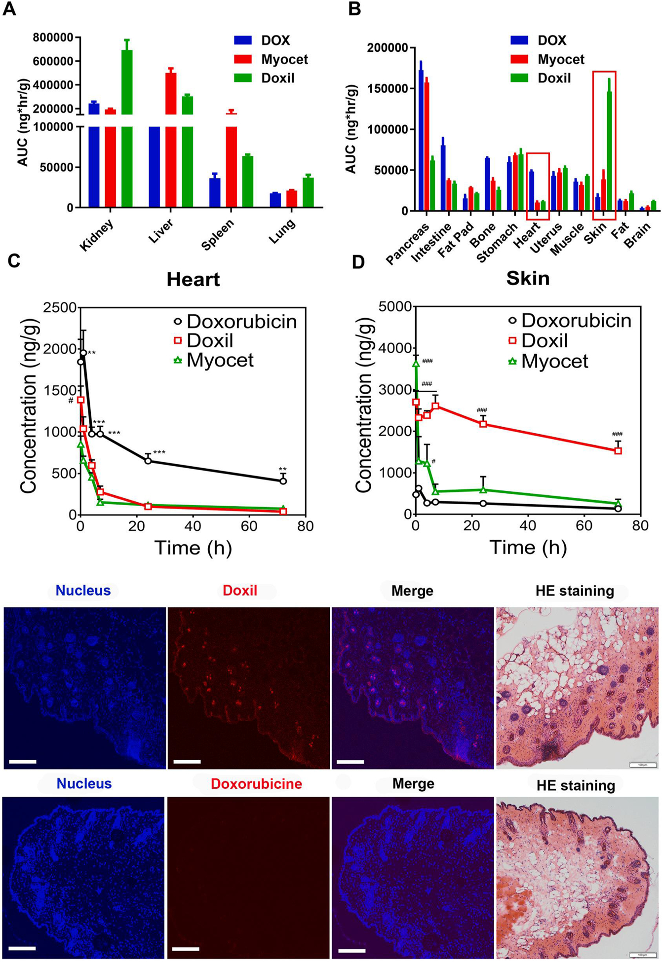
Long circulating and stable nanoformulations of doxorubicin alter tissue distribution differently in various organs. AUC0–72h of different DOX formulations in elimination related organs (A, B) and elimination non-related organs (C, D). Drug concentration in skin (A) and heart (B) in subcutaneous breast cancer model after IV dose of doxorubicin, Doxil, and Myocet (5 mg/kg). The statistical analysis results of Fig A and B are shown in supplementary materials (Table S4). #p < 0.01 DOX vs Myocet; **p < 0.01 DOX vs other two groups; ***p < 0.001 DOX vs other two groups; #p < 0.05 vs DOX; ###p < 0.001 vs DOX. (E) Confocal imaging to visualize detail localization of Doxil and doxorubicin in epidermis and dermis of the skin. Localization of Doxil throughout epidermis and dermis including hair follicles. Minimal presence of doxorubicin in epidermis and dermis tissues. Nuclear staining was performed using DAPI (Ex = 405 nm). The Doxil and doxorubicin were visualized at the Ex of 488 nm. Bar represents 100 μm H&E staining was performed in an adjacent section from series sections of the same tissue.