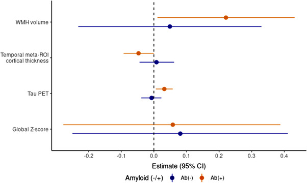
FIGURE 1.

Associations between plasma glial fibrillary acidic protein (GFAP) levels and cognitive and neuroimaging outcomes. Relationships between plasma GFAP levels and cognition and neuroimaging measures stratified by amyloid beta (Ab) status (elevated Ab PET, Ab[+]; non‐elevated Ab PET, Ab[–]). Amyloid and tau PET scores are log‐transformed. Coefficients are reflected for 100 pg/mL change in GFAP. WMH volume is expressed as a percentage of the total intercranial volume. CI, confidence interval; PET, positron emission tomography; ROI, region of interest; WMH, white matter hyperintensity
