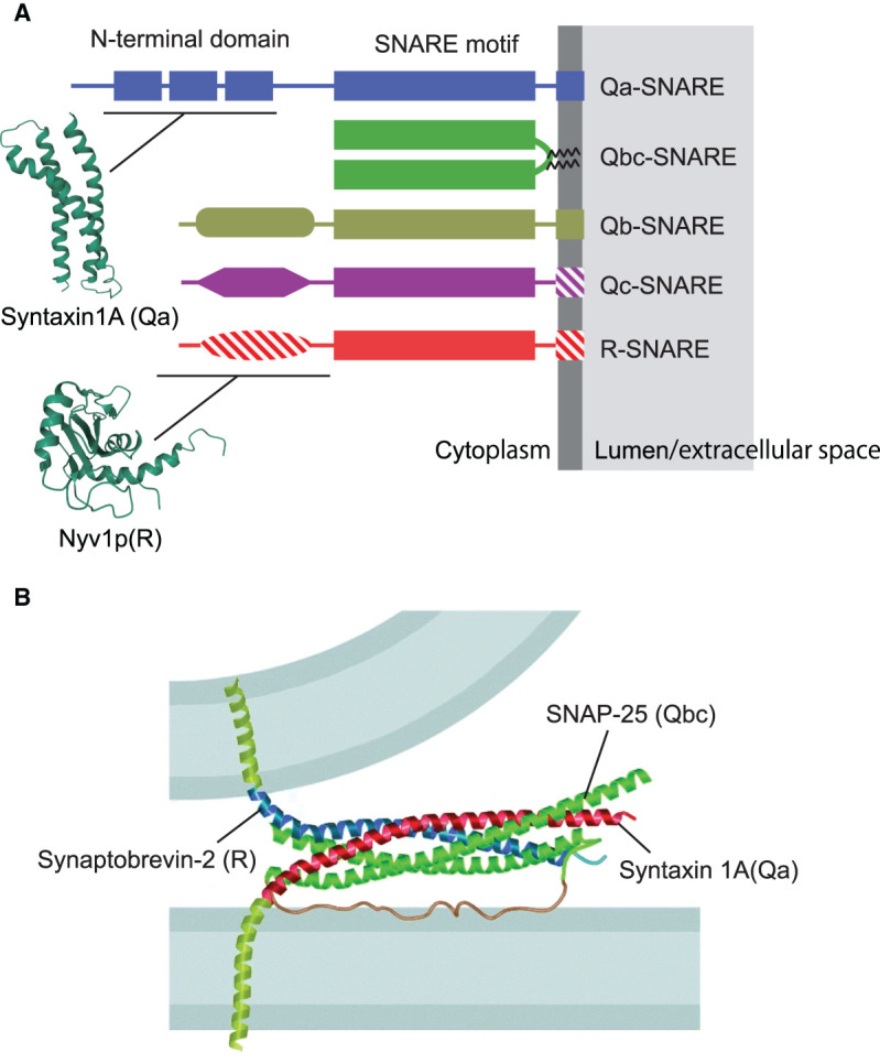
Figure 2. Domain structure of SNARE proteins.

(A) Overview over the main subfamilies of SNARE proteins Note that there is some variability within the subfamilies: Hatched areas signify domains that are absent from some of the family members. Moreover, transmembrane domains may be substituted by domains binding to PtdInxPx variants. The structures of the N-terminal domains of syntaxin 1A and Nyv1p are based on the Protein Data Bank entries 1S94 and 2FZ0, respectively (wwPDB.org). (B) Crystal structure of the neuronal SNARE complex, modeled between the two membranes destined to fuse. Note that the N-terminal region of syntaxin 1 was removed. Reproduced with permission from [24].
