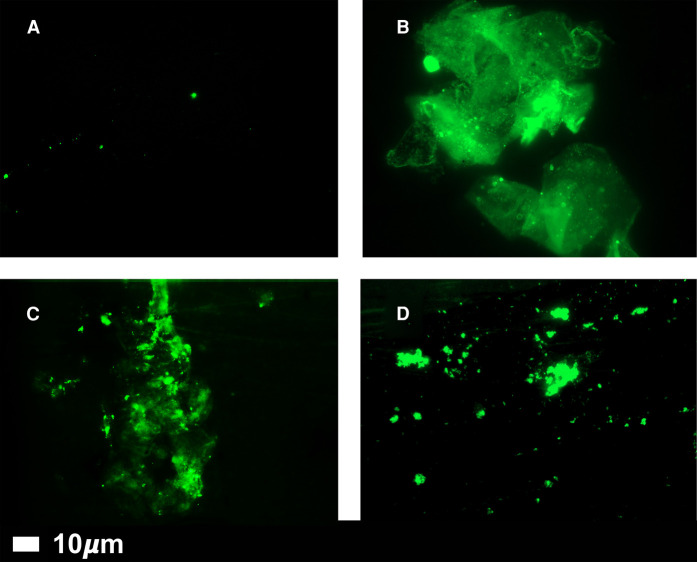
Figure 4. Fluorescence microscopy of representative micrographs showing microclots in the circulation of controls (A) and in patients with Long COVID (B–D).
Absence of significant amyloid microclots in the plasma of ‘normal’ individuals, and their significant presence in the plasma of individuals with long COVID. Platelet-poor plasma was produced by centrifugation at 3000×g for 15 min, stained with 5 µM thioflavin T, and imaged in a fluorescence microscope (Zeiss Axio Observer 7 with a Plan-Apochromat 63×/1.4 Oil DIC M27 objective (Carl Zeiss Microscopy, Munich, Germany). Wavelengths were Exc 450–488 nm/emission 499–529 nm, all as in [108].

