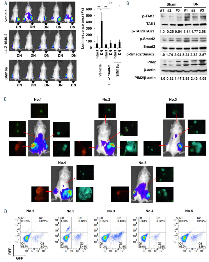
Figure 3.
Multiple myeloma tumor growth and dissemination upon immobilization. (A) Right and left hind legs in the same mice were subjected to sciatic denervation (DN) and sham operation (control), respectively. Two weeks later, luciferase-transfected mouse 5TGM1 multiple myeloma (MM) cells were simultaneously inoculated into tibiae in both immobilized (right) and intact (left) hind legs in the same mice. The TAK1 inhibitor LL-Z1640-2 or the PIM inhibitor SMI16a were intraperitoneally injected at 20 mg/kg twice a week. Control groups were given saline as a vehicle. IVIS images taken at 4 weeks. Tumor areas with luminescence shown in green, yellow and red were measured. Px: pixel. Data are expressed as the mean ± standard deviation (SD) (n=5). **P<0.01. (B) 5TGM1 MM cells were inoculated into right tibiae 2 weeks after DN or sham operation. The right tibiae with tumor lesions were harvested at 4 weeks after the MM cell inoculation. Cell lysates were then collected from MM tumor lesions, and protein levels of the indicated factors were analyzed by western blotting analysis. β- actin was used as a loading control. The following reagents were purchased from the indicated manufacturers: antibodies against phosphor-MAP3K7 (Thr187) from Cusabio (Cusabio Biotech, Wuhan, China); and antibodies against TAK1, PIM2, phosho-Smad2, Smad2, and β-actin from Cell Signaling Technology. (C) 5TGM1 MM cells transfected with green fluorescent protein (Gfp) or red fluorescent protein (Rfp) genes were inoculated into tibiae of right hind legs with DN and left sham-operated hind legs, respectively. The GFP-expressing 5TGM1/luc (5TGM1-GFP/Luc) cell line and the RFP-expressing 5TGM1/luc (5TGM1-RFP/Luc) cell line were generated by lentiviral transduction with the pLKO.1-puro-CMV-TurboGFP vector and pLKO.1-puro-CMV-TagRFP vector (Sigma-Aldrich, MO, USA), respectively. Four weeks later, IVIS images were taken. Blood was drawn from retro-orbital plexus, and tumors detected in the IVIS images were resected with surrounding tissues in the mice. Tumors emitting green or red fluorescence were visualized in resected samples with a fluorescence microscope (OLYMPUS SZX16). (D) Circulating 5TGM1-GFP and 5TGM1-RFP cells were analyzed in the blood samples from the mice at 4 weeks by flow cytometery.

