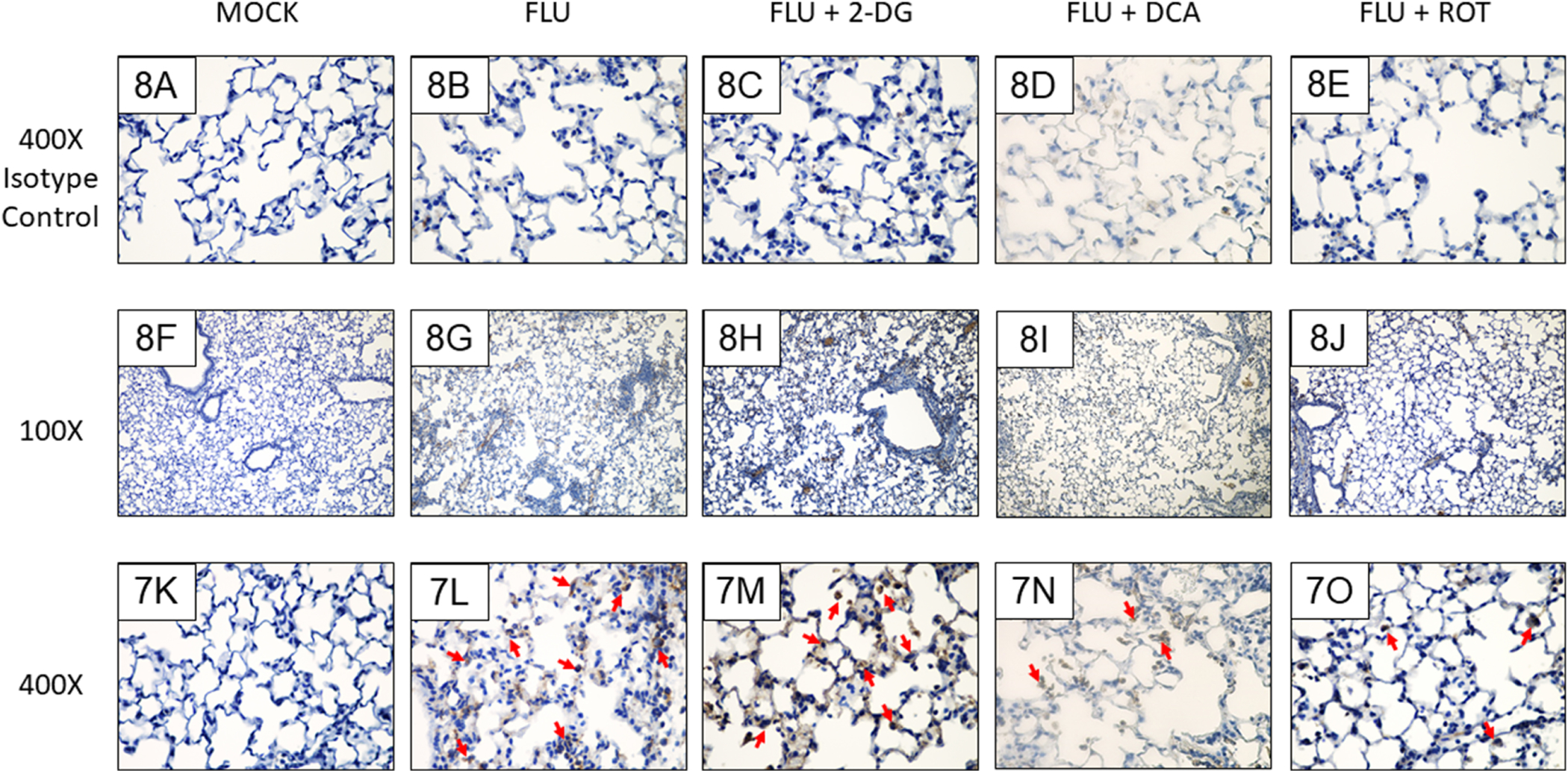
Figure 8. Both PDK and Complex I inhibition decrease alveolar epithelial 3-nitrotyrosine in IAV-infected mice.

Effect of mock and IAV infection (FLU) and treatment with 2-DG or ROT on day 6 3-nitrotyrosine formation in: (F and K) Mock-infected mice; (G and L) IAV-infected mice; (H and M) 2-DG-treated, IAV-infected mice; (I and N) DCA-treated, IAV-infected mice; and (J and O) ROT-treated, IAV-infected mice. (A-E) Isotype controls. All n = 3/group (from 2 independent mock or IAV infections per group). Magnifications, 400X (A-E and K-O), 100X (F-J). Representative photomicrographs are shown. Brightness and contrast were adjusted equally for all images to improve visibility. Red arrowheads indicate 3-nitrotyrosine-positive cells.
