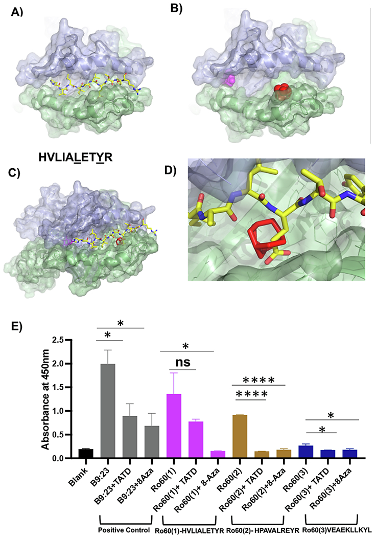
Fig. 1.

Structural modeling of a candidate autoantigen peptide and drug-like small molecules with I—Ag7 A) Ro60323-332HVLIALETYR, a peptide corresponding to murine Ro60, is shown as sticks modeled with I—Ag7 (PDB code 6BLX); yellow for carbon, blue for nitrogen, red for oxygen. The α-chain of I—Ag7 is shown in violet, β-chain in green. B) 8-Aza (magenta) and TATD (red) are shown as spheres in the top-scoring orientation posed by AutoDock Vina to the I—Ag7 antigen-binding cleft. C) Superposition of Ro60323-332HVLIALETYR on the I—Ag7 antigen-binding cleft in the presence of 8-azaguanine (magenta sticks) and TATD (red sticks). D) Steric clash between TATD and Ro60323-332HVLIALETYR provides a strategy to inhibit autoantigen peptide presentation. E) Measurement of IL-2 by absorbance. For consistency, the experiment was performed in triplicate and repeated twice. Splenocytes were stimulated with HVLIALETYR, HPAVALREYR, and VEAEKLLKYL for 24 h with and without TATD and 8-Aza. Cultured supernatants were collected to assay for IL-2 levels. The statistical differences between control and drug-injected mice were determined using a one-way ANOVA with an uncorrected Fisher’s LSD test at all threshold events. Error bars indicate with 95% class interval confidence, *: p < 0.0332, **: p < 0.0021, ***p < 0.0002, ****p < 0.0001, NS, not significant. (For interpretation of the references to colour in this figure legend, the reader is referred to the web version of this article.)
