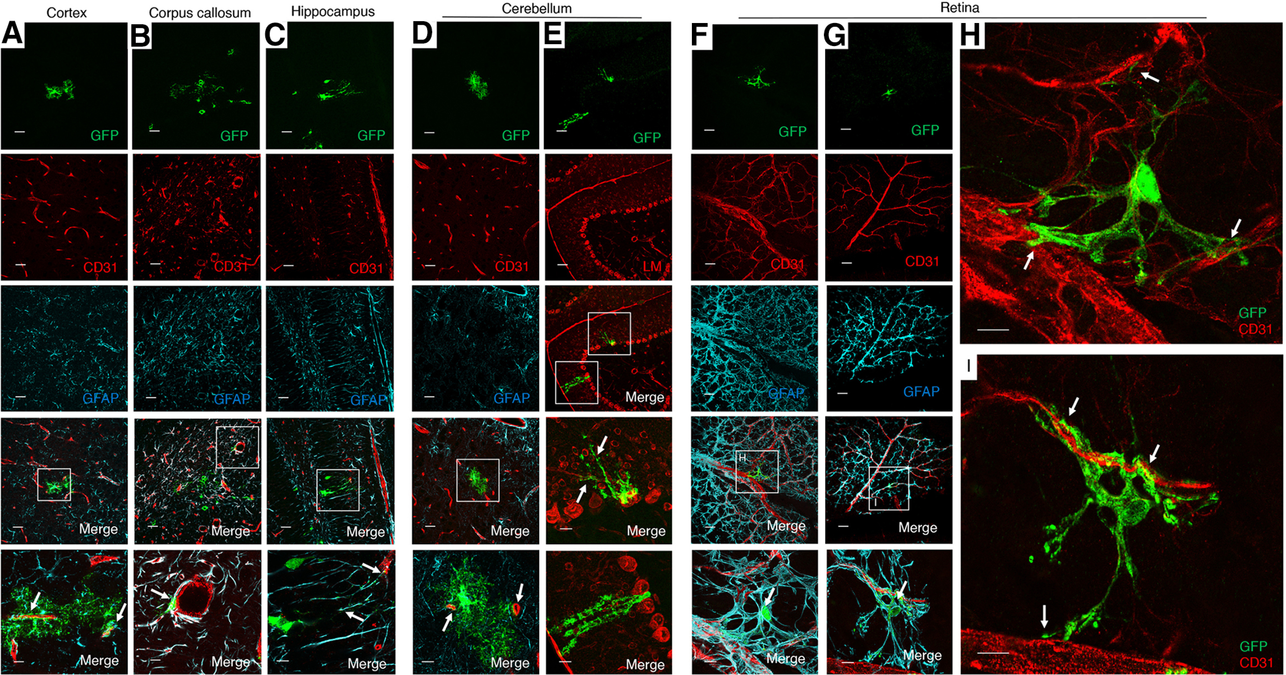
Figure 3.

Selective activation of MLCT in PAs of the P14 brain. A–D, MLCT;R26-YFP mouse pups were injected with tamoxifen from P1-P3, and brains were harvested at P14. Coronal sections were analyzed by triple immunofluorescence using anti-GFP (green), anti-CD31 (red), and anti-GFAP (cyan) antibodies. Note the coexpression of YFP and GFAP in some astrocytes of the cortex (A), corpus callosum (B), hippocampal dentate gyrus (C), and cerebellum (D). While many YFP+ PAs are also GFAP+, we detect some PAs that lack GFAP expression; for example, note the YFP+ PA in the cerebellar white matter that lack obvious GFAP expression (arrows in D). A–D, Bottom, Higher-magnification images of boxed areas in the lower-magnification merged panels. E, Representative image of a sagittal section through the MLCT;R26-YFP cerebellum at P30 analyzed by double immunofluorescence using anti-GFP (green) and anti-Laminin (red) antibodies. YFP+ Bergmann astrocytes are also GFAP+ (arrows), and Bergmann astrocyte cell bodies are located between Laminin+ Purkinje neuron cell bodies and extend elaborate processes (arrows). F, G, Flat-mounted retinas P14 MLCT;R26-YFP mice were analyzed by triple immunofluorescence using an anti-GFP antibody (green) in combination with anti-CD31 (red) and anti-GFAP antibodies (cyan). Note the overlapping YFP/GFAP expression in astrocytes of the P14 retinal primary vascular plexus (arrows in merged images, F and G). H, I, Higher-magnification images of boxed areas in F and G, respectively, showing only anti-GFP (green) and anti-CD31 (red) merged channels. Note the close juxtaposition between YFP+ PAs and CD31+ blood vessels (arrows). Scale bars, 100 µm.
