
Figure 14.
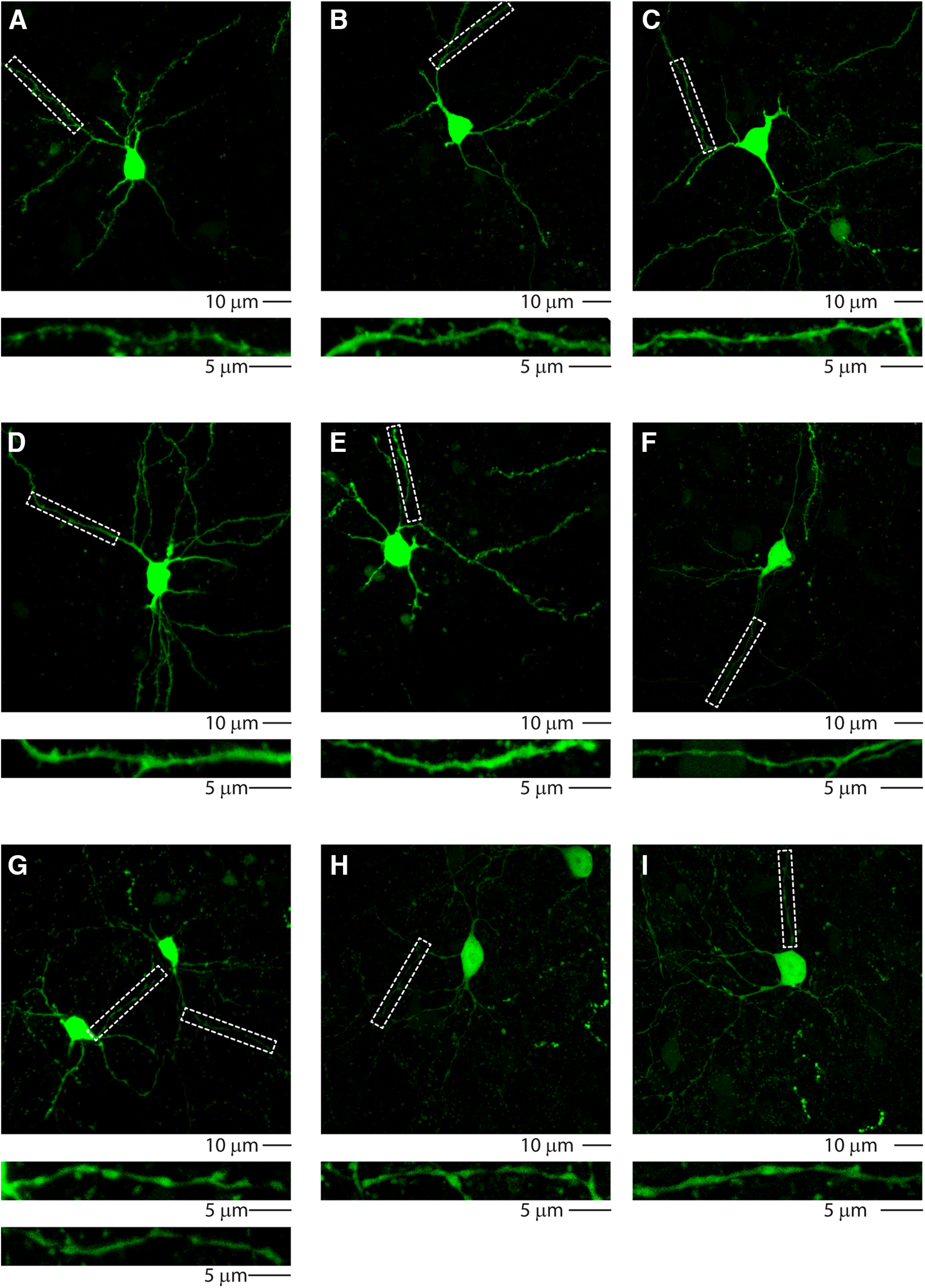
High-resolution images of intrastriatal Parv GABAergic neurons. High-resolution confocal images of biocytin-filled striatal neurons. Dashed lines indicate the corresponding enlargement of dendritic branches showing spiny (A–E) or smooth (F–I) morphology. Scale bars: low magnification, 10 µm; high magnification, 5 µm.