
FIGURE 2. High glucose differentially affects ion channel function in NHBE cells in vitro.
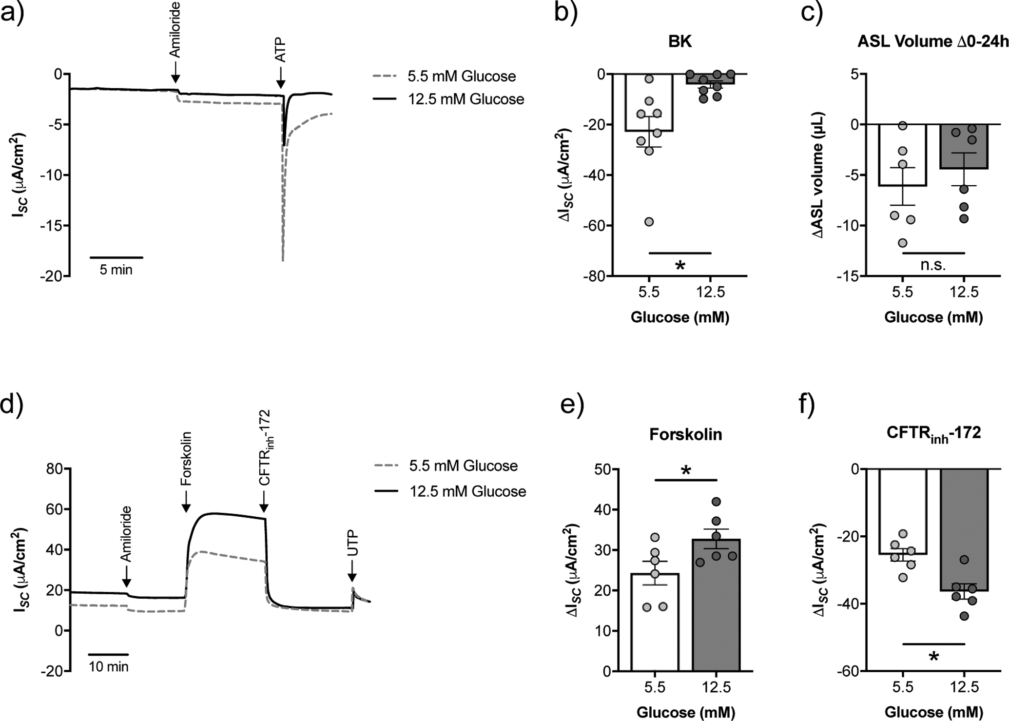
a) Representative tracing of ATP-stimulated ISC with a basolateral to apical K+ gradient [34] from fully differentiated NHBE cells cultured in ALI media with starting glucose concentrations of 5.5 mM or 12.5 mM. Ussing chamber experiments were performed on NHBE cells 20–24 h after basolateral media change. b) BK activity is significantly decreased in NHBE cells cultured under high glucose (n=8 from 6 lungs). c) Changes in ASL volume (ΔASL) over a 24 h period did not significantly differ between NHBE cells cultured under 5.5 mM or 12.5 mM glucose (n=6 from 3 lungs). d) Representative tracing of ISC following forskolin stimulation and CFTR inhibition by CFTRinh-172 from fully differentiated NHBE cells cultured in ALI media with 5.5 mM or 12.5 mM glucose. Ussing chamber experiments were performed on NHBE cells 20–24 h after basolateral media change. e) CFTR currents as measured by forskolin are significantly increased in NHBE cells cultured under high glucose (n=6 lungs). f) CFTR currents as measured by CFTRinh-172 inhibition are significantly increased in NHBE cells cultured under high glucose (n=6 lungs). * p < 0.05, Student’s t-test. Data are shown as mean ± S.E.M.