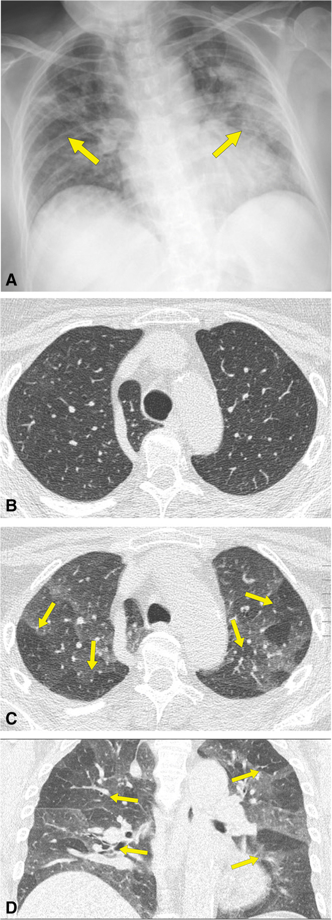
Figure 3.

A 67-year-old nonsmoking man who had COVID-19. A Anteroposterior chest radiograph shows bilateral and multifocal areas of consolidation (arrows). B Inspiratory CT scan at follow-up examination, 65 days after dyspnea and shortness of breath shows no abnormalities. C Expiratory CT scan, obtained at same level as B, and D coronal multiplanar reformation image shows significant bilateral patchy areas of low attenuation due to air trapping (arrows)
