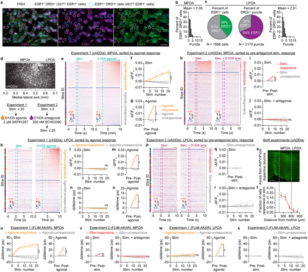
Extended Data Fig. 9 ∣. AVPV/PVpo dopamine neurons signal to Esr1 neurons in the MPOA through D1/D5 transmission.
a,b, Fluorescence in situ hybridization (using the RNAscope kit) demonstrates high co-localization of Drd1 and Esr1 expression in the MPOA (a), with an average of ~2 Drd1 puncta per Esr1 cell (n = 3 mice). The masks were selected based on Esr1 puncta. A cropped subregion of these images is shown in Fig. 4f. Scale bars: 100 μm.
c, In the LPOA, 39% of the Esr1 neurons also express Drd1 (average ~2 Drd1 puncta per Esr1 cell), and 68% of the Drd1 puncta are found in the Esr1 cells.
d, Sample field of view of cADDis expression in the Esr1 neurons in the MPOA (defined as the subregion of the preoptic area within 600 μm from the midline) and the LPOA (defined as the subregion beyond 600 μm from the midline). The definition of the boundary at 600 μm is derived from a standard mouse atlas62. Experimental designs are replotted here. In Experiment 1, we first used a concentric bipolar electrode to locally stimulate the AVPV/PVpo area for 20 rounds (20 Hz trains, 75 μA pulses for 1 s every 20 s) and then washed on the D1/D5 agonist SKF81297 (2 μM; diluted from 100 mM stock solution in dimethyl sulfoxide). In Experiment 2, we first used a minimal stimulation protocol to identify the MPOA Esr1 neurons that responded to AVPV/PVpo stimulations, washed on the D1/D5 antagonist SCH23390 (300 nM; diluted from 50 mM stock solution in saline), and then performed 20 more rounds of AVPV/PVpo stimulation in the presence of the antagonist.
e, Heatmaps showing the cell-by-cell cADDis response in the MPOA to both AVPV/PVpo stimulation (left) and D1/D5 agonist wash-on (right). Stimulation responses are averages of 20 trials, following baseline subtraction of the pre-stimulation means, while the agonist response is a single-trial response following baseline subtraction of the pre-agonist value. Esr1 neurons are divided into those that responded to the agonist (above the horizontal blue line) and those that did not (below the horizontal blue line) using a classifier (see Methods). Cells are sorted based on their agonist response and the same order is used for both panels. Gray lines on the top denote the windows that are used for quantification and classification. The color of the ‘Slide ID’ indicates the identity of the slice (n = 983 neurons from 4 slices, 3 mice) from which the cell was recorded.
f,g, In the agonist-responsive group (orange), neurons show persistent cAMP elevations (decreases in cADDis intensity) that accumulate after each AVPV/PVpo stimulation (f). The cAMP elevations are not seen in the agonist-unresponsive group (gray in f; n = 4 slices from 3 males). Panel g summarizes agonist responses (n = 4 slices from 3 males).
h, Heatmaps showing the cell-by-cell cADDis response of MPOA Esr1 neurons to AVPV/PVpo stimulation both before (left) and after washing on the D1/D5 antagonist (right). Stimulation responses are averages of 3 and 20 trials, following baseline subtraction of the pre-stimulation means. MPOA Esr1 neurons are divided into those that responded to the pre-antagonist stimulation (above the horizontal blue line) and those that did not (below the horizontal blue line) using a classifier (see Methods). Cells are sorted based on their stimulation response, and the same order is used for both panels. Gray lines on the top denote the windows that are used for quantification and classification. The color of the ‘Slide ID’ indicates the identity of the slice (n = 947 cells from 4 slices, 3 mice) from which the cell was recorded.
i,j, In the stimulation-responsive group (red), neurons show persistent cAMP elevations that are accumulated after each AVPV/PVpo stimulation (i: n = 4 slices from 3 males). The cAMP elevations are not seen after the antagonist wash-on (j: n = 4 slices from 3 males).
k, Same as e but for LPOA Esr1 neurons (n = 192 cells from 4 slices, 3 mice).
l,m, Same as f-g but for LPOA neurons. Note that LPOA Esr1 neurons (including those in the agonist-responsive group) did not respond to AVPV/PVpo stimulation (n = 4 slices from 3 males).
n,o, Same as l-m but for fluorescence lifetime measurements (a bleaching-insensitive way of analyzing fluorescence data; see Methods). Note that LPOA Esr1 neurons did not respond to AVPV/PVpo stimulation, including those in the agonist-responsive group (n = 4 slices from 3 males).
p-r, In Experiment 2, we again observed only very few AVPV/PVpo stimulation-responsive neurons in the LPOA, before or after antagonist application (n = 198 cells from 4 slices, 3 mice). Plotting conventions are the same as above. Note that we did not plot average responses of the stimulation-responsive group due to its small size.
s,t, In combined data from Experiments 1 and 2, the fraction of cells that responded to AVPV/PVpo stimulation gradually dropped off over distance from the midline (t; n = 2320 cells from 8 slices, 6 mice). Axon fields of AVPV/PVpo dopamine neurons shown here for comparison (s). Scale bars: 200 μm.
u, In the agonist-responsive group (orange), MPOA Esr1 neurons show persistent elevations in PKA activity (decreases in FLIM-AKAR lifetime) that accumulate across repeated trials of AVPV/PVpo stimulation. The cAMP elevations are not seen in the agonist-unresponsive group (gray; n = 4 slices from 3 males). The right panel summarizes agonist responses (n = 4 slices from 3 males).
v, In the stimulation-responsive group (red), MPOA Esr1 neurons show persistent elevations in PKA activity that accumulate after each AVPV/PVpo stimulation (left: n = 4 slices from 3 males). The elevations in PKA activity are not seen after antagonist wash-on (right: n = 4 slices from 3 males).
w, Same as u but for LPOA Esr1 neurons. Note that LPOA Esr1 neurons did not respond to AVPV/PVpo stimulation, including those in the agonist-responsive group (n = 4 slices from 3 males).
x, In Experiment 2, we again only recorded very few numbers of AVPV/PVpo stimulation-responsive Esr1 neurons in the LPOA, before or after antagonist. See above for plotting conventions. Note that we did not plot average responses of the stimulation-responsive group due to its small size.
Mean ± s.e.m. *p<0.05, **p<0.01, ***p<0.001. See Supplementary Table 1 for statistics.

