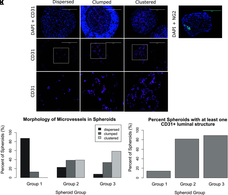
FIG. 2.
(A–I) Spheroids were categorized by morphology into three groups. Purple = CD31, blue = DAPI. Scale bar in (A–F): 200 μm. Image size for (G–I): 200 × 200 μm. (A, D, G) “Dispersed” spheroids have no or sparse and scattered CD31-positive cells. (B, E, H) “Clumped” spheroids have scattered clumps of CD31-positive cells and include CD31-positive lumina with thin walls. (C, F, I) “Clustered” spheroids have organized “lines” and/or clusters of CD31-positive cells. (J) Comparison of microvessel morphology between spheroids groups. (K) Quantification of spheroid groups with at least one CD31 positive luminal structure per group. (L) NG2 stain for pericytes in a Group 2 EVC spheroid. Green = NG2, blue = DAPI. Scale bar: 200 μm.

