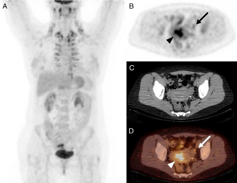
FIGURE 4.

18F-FDG PET/CT, A: MIP, B: PET, C: CT, and D: PET/CT, axial slice. Characterization and staging of newly diagnosed left ovarian mass complicated by constriction of left ureter and hydronephrosis in a 47-year-old woman (patient 17) with no history of endometriosis. Mildly increased, isolated 18F-FDG uptake in the peripheral part of cystoid left ovarian lesion (SUVmax 4.5, B and D: arrow) and increased 18F-FDG uptake in uterine cavity during menstrual flow (SUVmax 7.45, B and D: arrowhead). Left ovarian endometrioma was confirmed by histology.
