
Fig. 1.
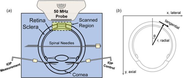
(a) Experimental setup of human donor globe inflation with high-frequency ultrasound imaging of the PPR, PPS, and ONH. (b) Illustration of the image scanning coordinate system (x/lateral, y/axial) and the polar coordinate system used to calculate radial and tangential strains.