. 2022 Feb 15;144(6):061001. doi: 10.1115/1.4053450

Fig. 3.

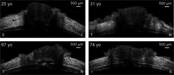
B-mode images of the posterior eye centered at the ONH from four human donors at the reference IOP (5 mmHg) showing the variability of the posterior eye tissue morphology across different age. The imaging axis is labeled for each eye. S: superior, I: inferior, N: nasal, T: temporal.

B-mode images of the posterior eye centered at the ONH from four human donors at the reference IOP (5 mmHg) showing the variability of the posterior eye tissue morphology across different age. The imaging axis is labeled for each eye. S: superior, I: inferior, N: nasal, T: temporal.